

Topics in the Chapter
- Introduction
- Meristematic Tissues
- Permanent Tissue
- Simple permanent tissue
- Difference between Parenchyma, Collenchyma and Sclerenchyma
- Complex Permanent tissues
- Xylem
- Phloem
- Epithelial Tissue
- Connective Tissue
- Fluid or vascular
- Skeletal Tissue
- connective Tissue
- Aerolar tissue
- Adipose tissue
- Muscular tissue
- Nervous tissue
Plant Tissues
Classification of Meristematic Tissues
- A group of cells with similar structure or function forms a tissue.
- Most plant tissues provide structural strength.
- These tissues are often dead, requiring less maintenance while providing mechanical strength.
- Plant tissues are categorized into:1.Meristematic tissues & 2. Permanent tissues
Meristematic Tissue
- Living tissue made of thin-walled, compact cells capable of division and growth.
Features of Meristematic Tissues:
- Thin cell walls made of cellulose.
- No intercellular spaces (cells are tightly packed).
- No vacuoles, but have dense cytoplasm and prominent nuclei.
- Contain a large number of cell organelles.
- In an active metabolic state, so they do not store food.
- Found in actively growing regions such as root and shoot tips.
Classification of Meristematic Tissues
1. Based on Origin:
Primary (Promeristem):
- Originates from the embryo meristem.
- Leads to the primary growth (height, length) of plants.
Secondary Meristematic Tissues:
- Formed by permanent tissues.
- Increases the diameter of plants, contributing to secondary growth.
2. Based on Location:
Apical Meristem:
- Located at the growing tips of stems and roots.
- Leads to the elongation of stems and roots (primary growth).
Intercalary Meristem:
- Located behind the apex.
- Part of the apical meristem, left behind during growth.
- Found at the base of leaves and internodes.
- Responsible for increasing leaf length (examples: grass, bamboo, mint).
Lateral Meristem:
- Also called secondary meristem.
- Found along the sides of the plant's longitudinal axis.
- Produces vascular tissues and causes an increase in girth.
- Responsible for secondary growth.

Permanent Tissues Overview
Types of Permanent Tissues
Simple Permanent Tissues
2. Supporting Tissues
(i) Parenchyma
- Permanent tissues consist of cells that lose the ability to divide.
- Cells have a definite shape, size, and thickness.
- Permanent tissues can be living or dead.
- They arise from meristematic tissues through cell division and differentiation (process where cells become specialized for specific functions).
- These cells have permanent shape, size, and function.
Types of Permanent Tissues
- Simple Permanent Tissues
- Complex Permanent Tissues
Simple Permanent Tissues
- Composed of cells that are similar in structure and function.
- Divided into: 1. Protective Tissues, & 2. Supporting Tissues
1. Protective Tissues
- These tissues serve a protective function.
- Include Epidermis and Cork (Phellem).
(i) Epidermis
- Forms a one-cell thick outer layer on plant organs (e.g., leaves, flowers, stems, roots).
- Covered by cuticle (waterproof waxy layer made of cutin).
- Thicker cuticle in xerophytes (plants in dry environments).
- Stomata (small pores) present in the epidermis for gas exchange.
- Guard cells (bean-shaped, with chloroplasts) surround the stomata.
Functions of Epidermis:
- Protects the plant from water loss and infection.
- Cuticle reduces transpiration (water evaporation) to prevent wilting.
- Stomata allow gas exchange (photosynthesis, respiration).
- Stomata help in transpiration.
(ii) Cork (Phellem)
- Found in older stems and roots.
- Composed of dead cells with thick walls.
- Cell walls contain suberin (waxy material), making them impermeable to water and gases.
- Cork cells lack protoplasm, filled with resins or tannins.
Functions of Cork:
- Acts as a protective barrier, preventing desiccation and injury.
- Cork is used commercially for its properties like lightness, toughness, elasticity, and compressibility.
- Applications: insulation, shock absorption, sports goods (cricket balls, shuttlecocks).
2. Supporting Tissues
- Provide mechanical support to the plant.
- Types of Supporting Tissues:1. Parenchyma, 2. Collenchyma & 3. Sclerenchyma
(i) Parenchyma
- Fundamental tissue found in plants.
- Thin-walled, oval or spherical cells.
- Cell walls composed of cellulose and pectin.
- Contains a large central vacuole for food and water storage.
- Primary function: storage of food.
- Idioblasts are parenchyma cells that store resins, tannins, gums, oils.
- Chlorenchyma (parenchyma with chloroplasts) performs photosynthesis (e.g., in mesophyll of leaves).
- Aerenchyma (parenchyma with air spaces) in aquatic plants provides buoyancy.
- Parenchyma provides turgidity (firmness) to cells.
(ii) Collenchyma
- Living mechanical tissue.
- Elongated cells with thickened corners (due to cellulose and pectin).
- Provides flexibility to plant parts, allowing bending without breaking.
- Found in herbaceous dicot stems and leaf margins.
- Provides mechanical strength and elasticity to growing stems.
- May contain a few chloroplasts.
(iii) Sclerenchyma
- Strengthening tissue, composed of dead cells with thick lignified walls.
- Lignin makes the cells waterproof.
- No intercellular spaces.
- Two types of sclerenchyma cells:
- Small cells with very thick walls and tiny lumens.
- Found in drupes (e.g., mango, coconut), legume seeds.
- Long, narrow, and lignified cells.
- Lumens are larger than in sclereids.
- Used commercially in making ropes, mats, and textile fibers (e.g., jute, coir).

Difference between Parenchyma, Collenchyma, and Sclerenchyma

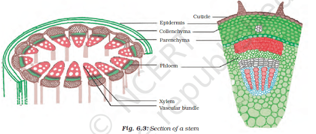
Complex Permanent Tissues: Xylem & Phloem
- Complex permanent tissues have more than one cell type that work together.
- Responsible for transport of organic materials, water, and minerals.
- These tissues form the vascular bundles (conducting tissues).

Xylem
- Also called wood, xylem is a vascular tissue responsible for water and mineral conduction.
- Tracheids: Elongated, dead cells involved in water conduction (found in gymnosperms).
- Vessels: Cylindrical tubes forming a continuous channel (found in angiosperms).
- Xylem Parenchyma: Stores starch (food).
- Xylem Sclerenchyma: Non-living fibres providing mechanical support.
- Annual xylem rings in tree trunks help determine the age of the tree.
Phloem
- Phloem conducts food (sugars) in both directions within the plant.
- Sieve Tubes: Elongated cells with sieve plates for food conduction.
- Companion Cells: Assist sieve tubes with metabolic activities; sister cells to sieve tubes.
- Phloem Fibres: Provide mechanical support.
- Phloem Parenchyma: Stores food and aids in radial conduction.
- In xylem, movement is unidirectional, but in phloem, it is bidirectional.
Comparison Between Xylem and Phloem

- Connective tissue: Widely spaced cells embedded in an intercellular matrix.
- Matrix composition determines the tissue's function.
- Fibres: Contains white and yellow fibres.
- Primary function: Provides support and helps keep organs in place.
Types of Connective Tissue
1. Fluid or Vascular Tissue (blood and lymph)
Blood:
- Matrix: Fluid matrix is called plasma.
- Functions: Transports nutrients, gases, excretory products, and hormones.
Plasma:
- Makes up 55% of blood.
- Consists of 90-91% water, 7% proteins (Albumin, fibrinogen, globulin), and 0.9% inorganic salts.
Corpuscles:
- Make up 45% of blood.
- Red Blood Cells (RBCs): Contain haemoglobin, a red pigment that helps in oxygen transportation.
- White Blood Cells (WBCs): Also called the "soldiers of the body."
- Function: Protects the body by engulfing bacteria and foreign particles.
- Types of WBCs: Monocytes, Lymphocytes, Basophils, Neutrophils, Eosinophils.
- Blood Platelets (Thrombocytes):
- Spindle-shaped cells involved in blood clotting.
2. Skeletal Tissue
- Hard connective tissue that forms the body’s supportive framework.
- Types: Bone and Cartilage.
Bone:
- Matrix: Hard due to calcium phosphate, CaCO3 (60-70%), and a protein called ossein.
- Bone cells (osteoblasts): Embedded in the hard matrix.
- Lamellae: Concentric layers of matrix around a central canal.
- Marrow cavity: Hollow space in long bones filled with bone marrow.
Cartilage:
- Elastic and less hard compared to bones.
- Protein chondrin provides elasticity.
- Chondroblasts: Widely spaced cells in a matrix reinforced by fibres.
- Locations: Joints, nose, ear, trachea, larynx.
- Provides flexibility and tensile strength.
3. Fibrous Connective Tissue
- Divided into Yellow fibrous connective tissue and White fibrous connective tissue.
Yellow fibrous connective tissue:
- Elastic due to yellow fibres in the matrix.
- Forms ligaments that connect bone to bone.
White fibrous connective tissue:
- Contains white fibres with little matrix.
- Forms tendons that attach muscles to bones.
4. Areolar Tissue
- The most widely distributed connective tissue in the body.
- Fills spaces inside organs.
- Found between skin and muscles, around blood vessels, nerves, and in bone marrow.
5. Adipose Tissue
- Oval/round cells filled with fat globules called adipocytes.
- Locations: Found beneath the skin, around the heart, brain, and under eyeballs.
- Functions: Acts as an insulator and prevents heat loss.
Muscular Tissue: Overview
- Muscular tissues enable movement in the body.
- Made up of long, fibre-like cells known as muscle fibres.
- These fibres can contract and relax.
- Types of Muscular Tissue:1.Striated muscles, 2.Cardiac muscle fibres, 3.Non-striated muscles
Types of Muscular Tissue
1. Striated Muscles (Voluntary Muscles)
- Also called voluntary muscles since they are controlled consciously.
- Muscle fibres are multinucleated and unbranched.
- Enclosed by a thin membrane called sarcolemma.
- Cytoplasm of these fibres is called sarcoplasm.
- These muscles tire easily and require rest.
2. Cardiac Muscle Fibres
- Involuntary muscles found only in the heart walls.
- Structure is between striated and non-striated muscles.
- Uninucleated and branched fibres, connected by intercalated discs.
- Responsible for rhythmic contraction and relaxation throughout life.
3. Non-striated Muscles (Smooth Muscles)
- Also called involuntary muscles.
- Uninucleated, spindle-shaped fibres.
- Not enclosed by a membrane, but fibres join together in bundles.
- Found in organs like the stomach, intestine, urinary bladder, bronchi, and iris of the eye.
- Responsible for peristaltic movements in the alimentary canal.
Nervous Tissue: Overview
- Specialized tissue enabling animals to perceive and respond to stimuli.
- The functional unit is the neuron or nerve cell.
- The cell body is called cyton and is covered by a plasma membrane.

Neuron Structure
1. Dendron:
- Short, hair-like extensions from the cyton.
- Subdivided into dendrites.
2. Axon:
- A long, cylindrical process extending from the neuron, with fine branches at the end.
- Covered by a sheath.
- Transmits impulses between neurons via electrochemical waves.
- The point where an axon of one neuron connects closely to the dendrites of another neuron is called a synapse.
0 Comments